ctbrazerzkidai.blogg.se

Cephalic flexture
Cephalic flexture


cephalic flexture

  • optic pits begin to project laterally (diencephalic floor has expanded).
  • maxillary process begins to differentiate.
  • head fold in amnion, to about level of otic placode.
  • no closure of neural fold anterior to myelencephalon.
  • spread of neural crest into frontonasal processes.
  • first contact of neural folds closed up to otic capsule.
  • robust mass of mesenchyme/neural crest in future 1 st arch/frontonasal region.
  • forebrain = diencephalon only (identified by optic pit) – thin flap projection anteriorly on embryo.
  • optic pits and otic placode visible optic pits rounded.
  • first appearance of optic pit (slit like).
  • first arch mesenchyme condensing into recognizable mass.
  • See data on age/stage relations at end of series. Ages are mean age, calculated from time of mating.
  • wiki-commons:Special:FilePath/Isthmic_Organizer_TFs_and_signaling_molecules.All photographs taken immediately after removing embryo from uterus, with no preparation.
  • wiki-commons:Special:FilePath/Gray651.png STAGE 26 definitive cephalic flexure no closure of neural fold anterior to myelencephalon head fold in amnion, to about level of otic placode forelimb. abnormal cerebellum development abnormal pontine flexure morphology abnormal rhombomere morphology (47 genotypes, 75 annotations).
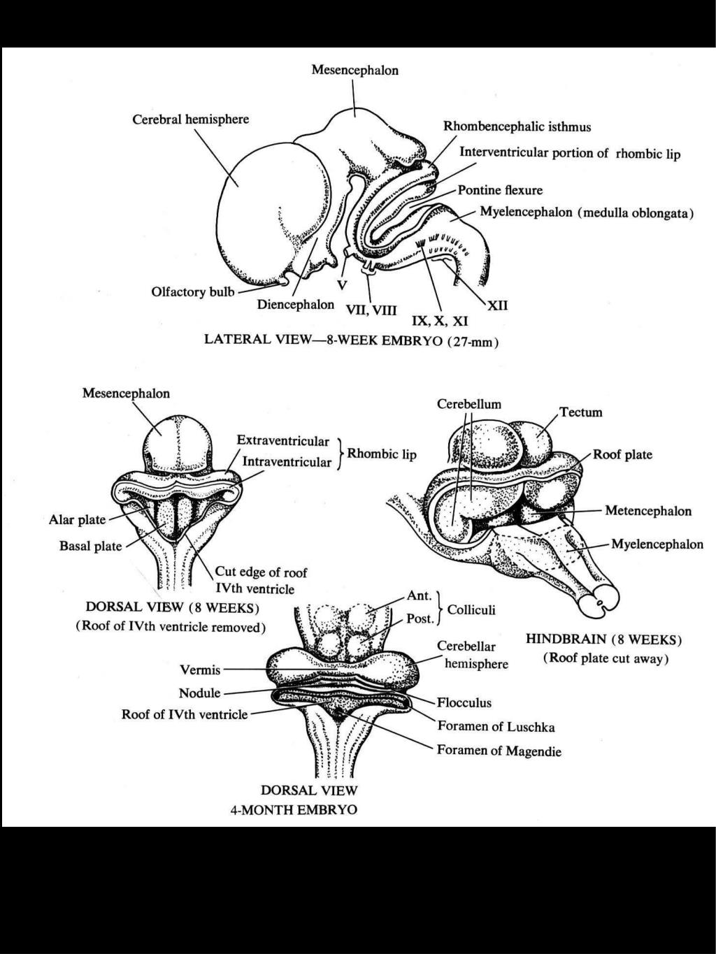
  • Brain of human embryo of four and a half weeks, showing the three flexures (en).
  • It also develops by the 5th week of gestation. The cervical flexure bends the neuraxis between the brain and the spinal cord. This flexure develops by the 5th week of gestation.
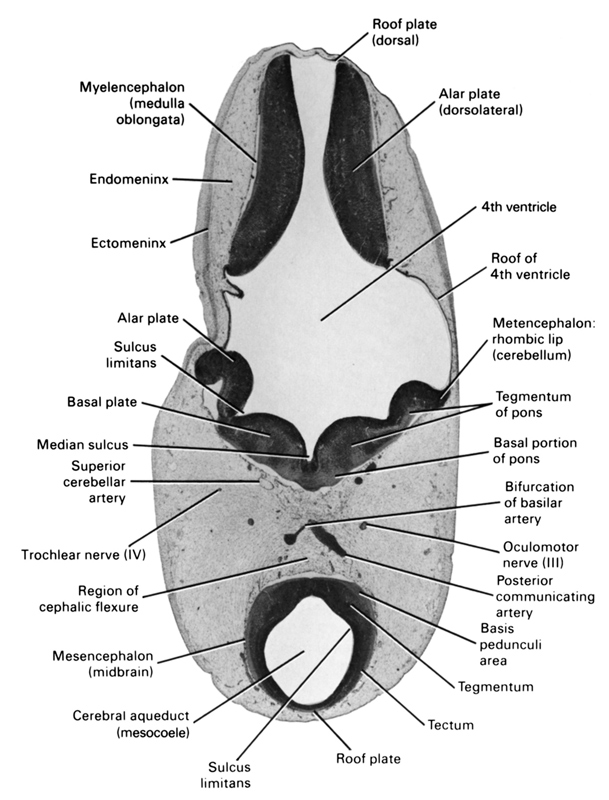
  • dbr:File:Isthmic_Organizer_TFs_and_signaling_molecules.png The cephalic flexure (also known as the midbrain flexure) bends the neuraxis at the junction of the midbrain and hindbrain.
  • wiki-commons:Special:FilePath/Gray651.png?width=300.
  • These flexures have formed by the time that the primary brain vesicles have developed into five secondary brain vesicles in the fifth week. It also bends dorsally into the pontine flexure. This limitation causes the neural tube to bend, or flex, at two ventral flexures – the rostral cephalic flexure, and the caudal cervical flexure.

    cephalic flexture

    The space into which the cranial part of the neural tube is developing is limited.
    cephalic flexture

    Pages 44 This preview shows page 20 - 38 out of 44 pages. School University of New South Wales Course Title ANAT 2341 Type. Early development of the cns cephalic flexure. At four weeks gestational age in the human embryo the neural tube has developed at the cranial end into three swellings – the primary brain vesicles. Early development of the CNS Cephalic flexure Cervical flexure 3 primary brain. Three flexures form in the part of the embryonic neural tube that develops into the brain.


    Cephalic flexture